The Science

DNA Curtain Technology

A nanofabrication-based approach that transforms single-molecule biophysics from a low-throughput niche technique into a broadly accessible, high-throughput research tool.

The Problem

DNA-protein interactions are hard to study at scale

DNA-protein interactions — including replication, transcription, and repair — are essential for cell survival and lie at the root of many genetic diseases. Understanding them at the molecular level is fundamental to biology and medicine.

Traditional bulk (ensemble) techniques average over millions of molecules, masking hidden subpopulations and transient conformational states. Single-molecule methods reveal these hidden dynamics but have historically suffered from low throughput and complex, indirect data interpretation.

As a result, powerful single-molecule assays have remained largely inaccessible — confined to highly specialized biophysical laboratories.

Bulk vs. Single-Molecule

Bulk / Ensemble
Averages over populations. Fast and quantitative, but cannot resolve subpopulations, transient states, or dynamic heterogeneity.
Traditional Single-Molecule
Reveals hidden states. But low throughput, indirect probing, and data interpretation demands specialized expertise.
DNA Curtains
100+ molecules in parallel. Direct, real-time visualization. Statistically robust data from a single experiment. Accessible to any TIRF lab.
The Solution

How DNA Curtains work

Nanofabricated barriers on a glass surface, combined with a supported lipid bilayer and gentle flow, create an ordered array of individually addressable DNA molecules ready for fluorescence imaging.

Step 1 of 3

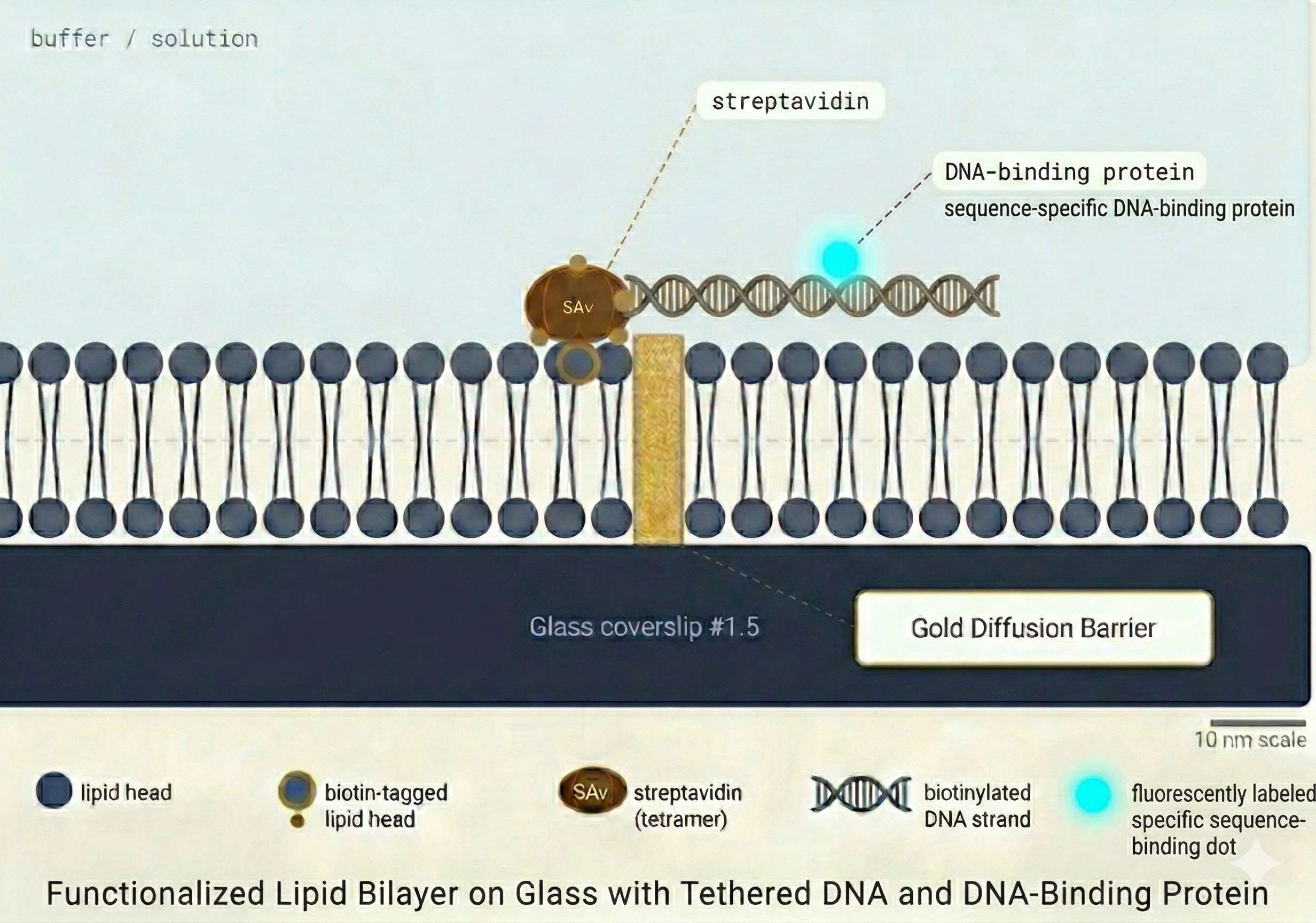
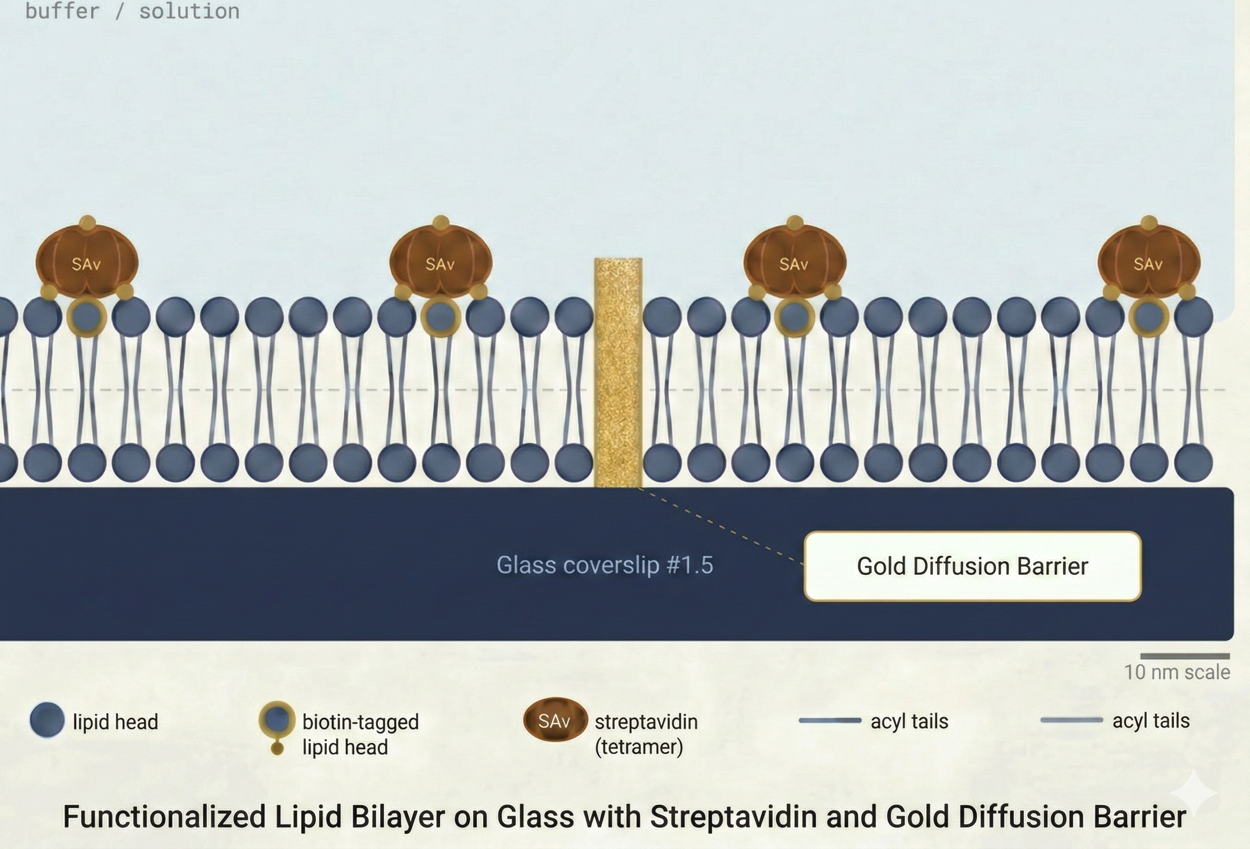
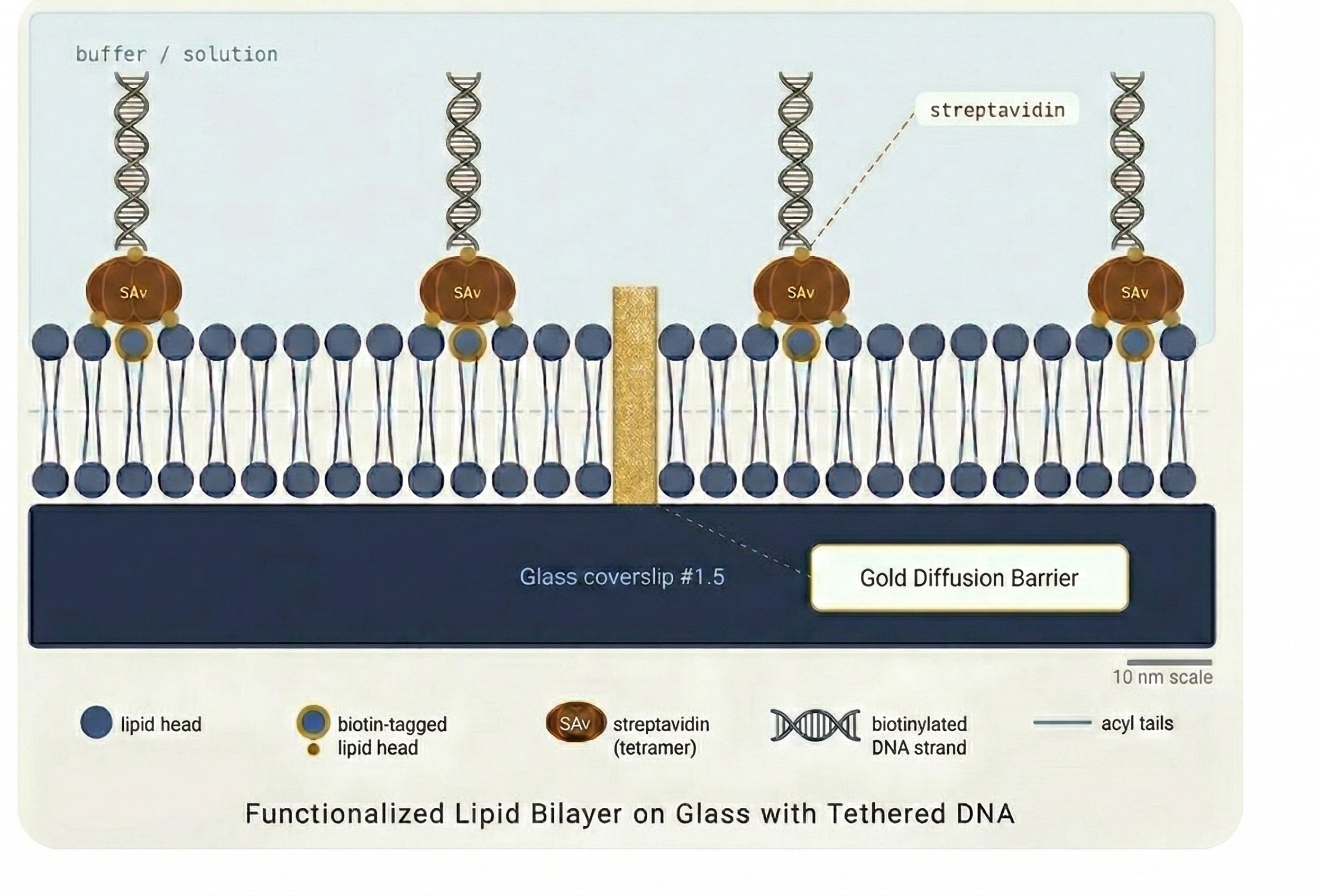
Lipid Bilayer Formation

Before any DNA experiment, all surfaces of the microfluidic flow channel are passivated using a supported lipid bilayer (SLB). The SLB serves two critical functions:

  • Mobility: Lipid-anchored DNA molecules can diffuse along the bilayer surface, allowing them to migrate toward barriers under flow.
  • Passivation: The SLB minimizes non-specific adsorption of proteins, reducing background fluorescence and ensuring clean data.

The bilayer is formed directly within the flow cell, using standard vesicle fusion protocols compatible with most lipid compositions.

Functionalized lipid bilayer on glass coverslip with streptavidin and gold diffusion barrier
Step 2 of 3

DNA Tethering

DNA molecules (typically 20–50 kbp in length) are tethered to the supported lipid bilayer via a biotin–streptavidin interaction at one end (single tether) or both ends (double tether).

Once introduced into the flow cell, a small and controlled flow is applied. Because the DNA is anchored to the mobile bilayer, the hydrodynamic drag extends and drives the DNA molecules toward the nanofabricated barriers — without stretching or damaging them.

DNA molecules tethered to the lipid bilayer via biotin-streptavidin, extended by hydrodynamic flow toward the nanofabricated barrier
Step 3 of 3

Curtain Formation & Imaging

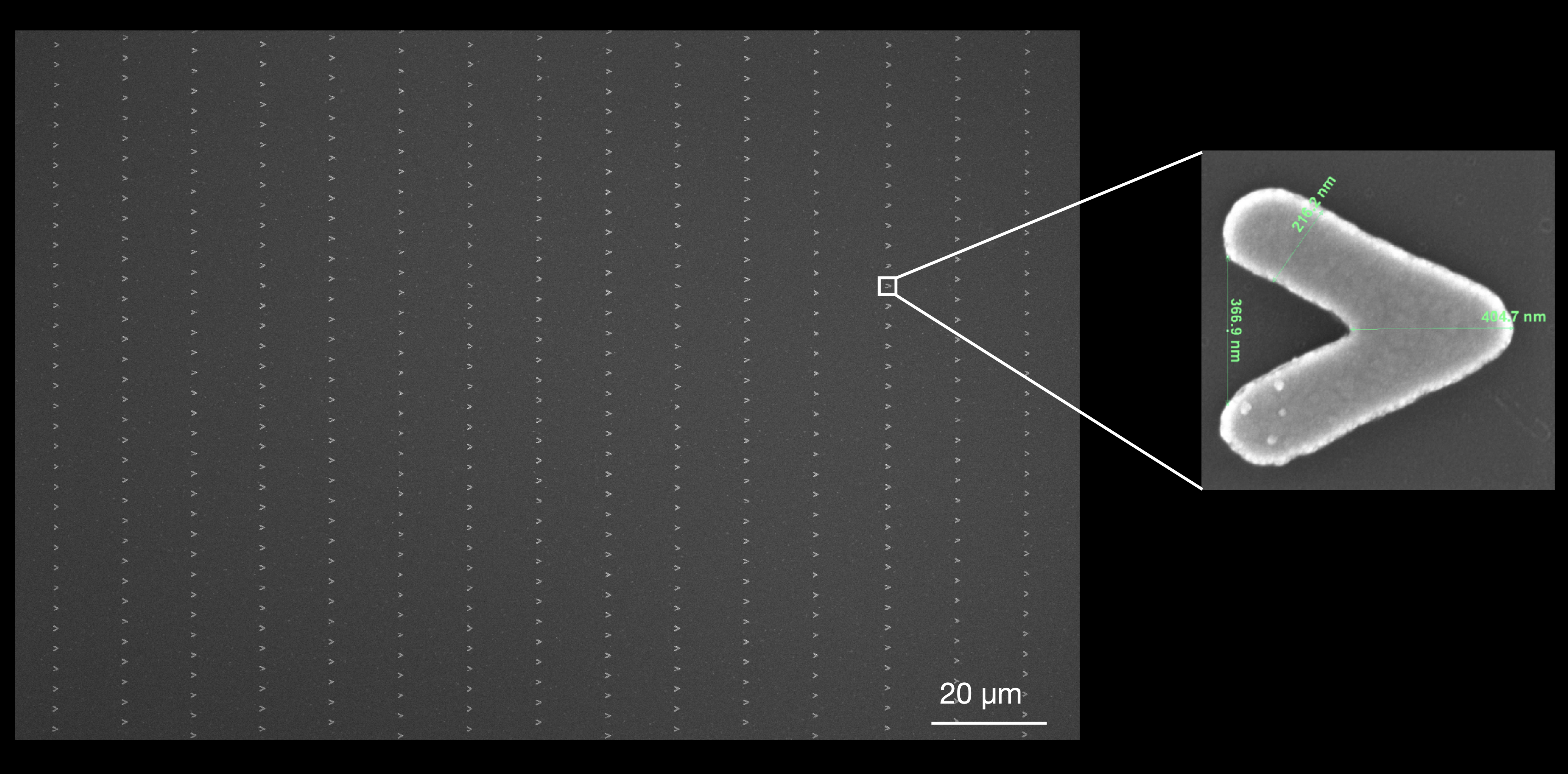
When DNA molecules reach the nanofabricated barriers, the barriers physically capture them at their leading edges. This forms the DNA Curtain — an ordered, parallel array of individually extended and spatially separated DNA molecules, all aligned along the barrier edge.

With flow maintained, the DNA remain extended and accessible. Fluorescently labeled proteins of interest are then introduced, and their binding, diffusion, and activity along individual DNA molecules is visualized in real time using objective-type TIRF microscopy.

Automated tracking scripts extract kymographs for every molecule in the field of view simultaneously, generating statistically robust, quantitative data on diffusion coefficients, dwell times, processivity, and more — from a single experiment.

DNA Curtain — aligned DNA molecules at barrier with fluorescent protein spots, TIRF evanescent field, and kymograph inset
Nanofabrication

Precision-engineered barriers

The performance of a DNA Curtain depends critically on the quality and geometry of its nanofabricated barriers. 1NA's expertise in electron-beam lithography enables barrier designs not achievable with conventional approaches.

SEM image of V-shaped nanofabricated barriers
SEM image of 1NA's v-shaped barrier array (left) and close-up of a single barrier with dimensions (right). Scale bar: 10 µm.
1NA Unique Design

V-Shaped Barriers

1NA's proprietary v-shaped barrier design deliberately spaces out individual DNA molecules, ensuring each molecule is well-separated from its neighbor. This geometric spacing enables straightforward automated data analysis with minimal signal overlap between adjacent molecules.

Classic Design

Zigzag Continuous Barriers

The more traditional continuous zigzag barrier design, perfected by the Greene lab, is also available. This design captures DNA at the leading edges of continuous barrier lines and is well-established in the published literature.

Available Configurations

Tethering Mode
Single-tether (one-end anchored)
Double-tether (both ends anchored)
Barrier Material
Gold (Au)
Titanium (Ti)
Barrier Geometry
V-shaped (1NA design)
Zigzag continuous (Greene lab design)
Data Output

From raw imaging to quantitative insights

Kymographs

Time-position representations extracted from individual DNA molecules show protein movement, binding, and release events with exceptional clarity. Hundreds of kymographs are generated in parallel from a single experiment.

Diffusion Coefficients

Mean Square Displacement (MSD) analysis of tracked protein trajectories yields diffusion coefficients. Population histograms reveal heterogeneity in protein mobility not accessible to bulk methods.

Dwell Times & Dynamics

Binding dwell time distributions, velocity histograms, processivity measurements, and DNA compaction rates are extracted automatically, providing mechanistic insight into protein function.

Ready to Start?

Bring DNA Curtains technology to your lab

Compatible with any objective-type TIRF microscope. Contact us to discuss your setup and research application.

Contact Us View Product Specs