DNA Curtains technology has been applied across core areas of molecular biology, enabling discoveries at the intersection of DNA biology, protein biochemistry, and genome maintenance.
DNA double-strand breaks are among the most cytotoxic lesions a cell can sustain. Understanding how repair complexes recognize, process, and fix these breaks is fundamental to cancer biology, aging research, and genome stability.
DNA Curtains enable direct, real-time observation of repair machinery in action. The MRN complex (Mre11-Rad50-Nbs1), BLM helicase, CtIP, and other key repair factors have been visualized on individual DNA substrates — revealing recruitment dynamics, resection mechanisms, and pathway choice at the single-molecule level.
Fluorescent labeling of specific protein components combined with multi-color TIRF imaging allows simultaneous tracking of multiple repair factors, revealing their spatiotemporal coordination.
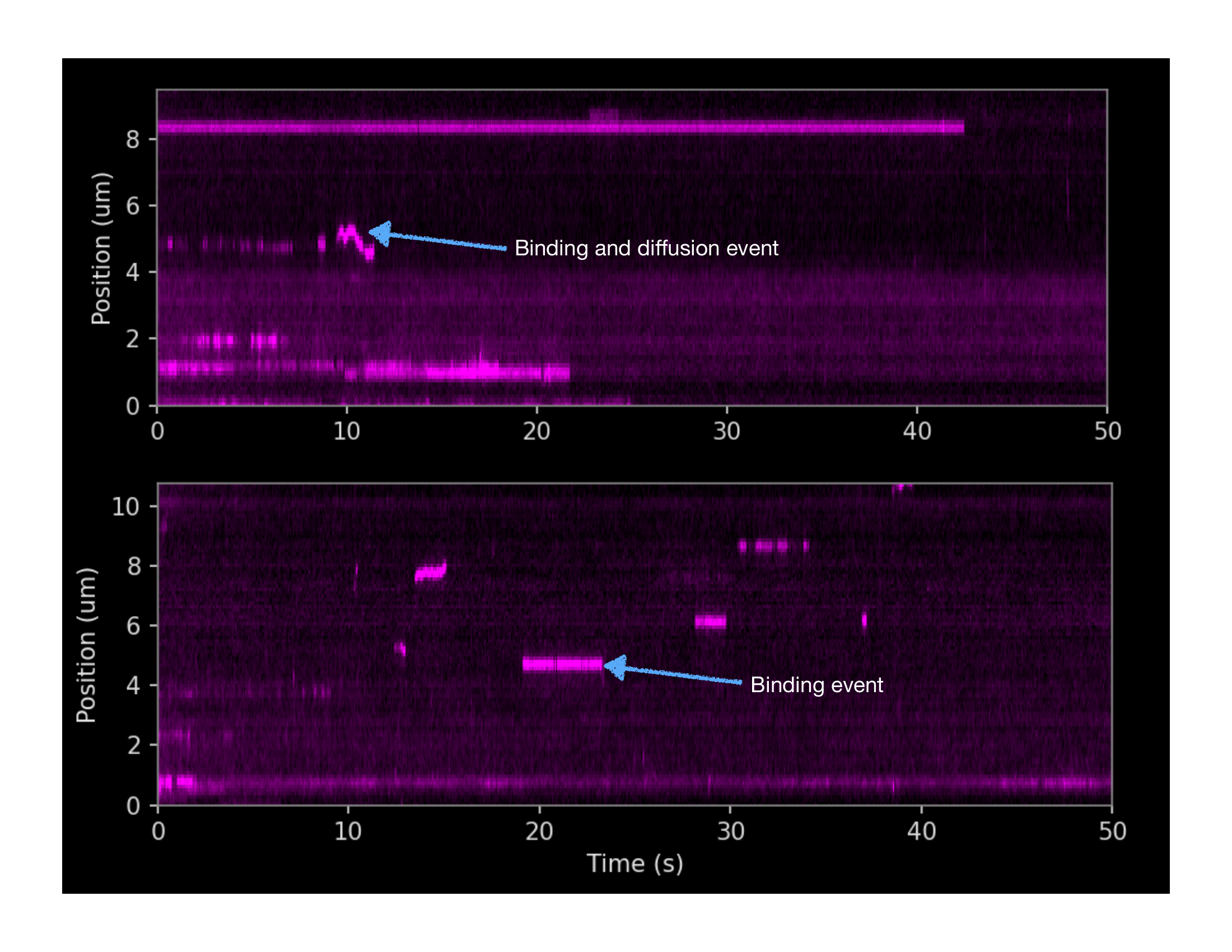
Binding kinetics, and diffusion of DNA-binding proteins are easily visualized as kymographs by picking out an individual DNA tether out of hundreds recorded, and plotting protein binding position along the tether against time.
Both panels on the left show the binding behavior of proteins to one individual DNA tether. Dwell times, diffusion behavior and binding location along the DNA strand can be visualized and quantified.
Helicases, translocases, polymerases, and other DNA motor proteins move along DNA in an ATP-dependent manner. Their velocity, processivity, and response to roadblocks are critical parameters for understanding their cellular functions.
DNA Curtains provide an ideal platform for tracking motor protein dynamics. Individual molecules are simultaneously imaged as they translocate along hundreds of DNA tethers, generating statistically rich datasets in a single experiment.
Quantitative parameters extracted from kymograph analysis — including velocity distributions, MSD curves, and diffusion coefficients — reveal mechanistic heterogeneity that would be entirely invisible to bulk measurements.
Directly visualize individual DNA-protein interactions with statistical robustness and intuitive data interpretation in a single experiment. High-throughput, Cost-effective, Innovative. That's DNA Curtain Technology.
The figure shows simulated DNA curtain data illustrating the movement of an arbitrary DNA translocase along DNA. From a single DNA tether, a kymograph is extracted to visualize protein movement along the DNA over time. Automated scripting allows simultaneous tracking of all DNA strands in the field of view, producing statistically robust, high-throughput data from a single experiment. Quantitative analysis allows users to extract dynamic parameters, such as Means Square Displacement and diffusion coefficients, binding dwell times, and more. Furthermore, DNA curtains enable direct observation of DNA-binding protein dynamics, unlike many other single-molecule techniques, making data interpretation not only more intuitive but also less dependent on complex data processing.
How proteins find their target sequences on genomic DNA is a fundamental question in biology. DNA Curtains are uniquely suited to studying the target search mechanisms of DNA-binding proteins — including nucleases, transcription factors, and CRISPR-Cas systems.
By directly watching individual protein molecules as they scan along DNA, DNA Curtains reveal whether a protein uses 3D diffusion, 1D sliding, or a combination of both. Cleavage events can be observed in real time as DNA shortening or strand release.
The CRISPR-Cas9 system has been studied extensively with DNA Curtains, revealing its PAM-proximal interrogation mechanism and the kinetics of R-loop formation and cleavage.
Sternberg et al. (2014) used DNA Curtains to visualize how Cas9 interrogates DNA for its target sequence. The work revealed that Cas9 uses 3D diffusion punctuated by brief interactions with PAM sequences to find its target — a foundational insight for CRISPR tool development.
Further work by Dillard et al. (2025) used DNA Curtains to characterize Cas9 inhibition by anti-CRISPR proteins, demonstrating the platform's continued relevance for cutting-edge CRISPR research.
How genomes are packaged, organized, and dynamically regulated is central to gene expression, cell identity, and disease. DNA Curtains have been applied to study nucleosome assembly and remodeling, cohesin-mediated loop extrusion, and CTCF-mediated genome architecture.
The ability to directly observe protein-driven DNA compaction, looping, and sliding in real time — with statistical power from hundreds of simultaneous events — makes DNA Curtains an exceptionally powerful tool for chromatin biology.
Loop extrusion by cohesin has been directly visualized using DNA Curtains (Kim et al., Science 2019), providing compelling evidence for the loop extrusion model of genome folding that now underpins our understanding of topologically associating domains (TADs).
Kim et al. directly visualized human cohesin compacting DNA by loop extrusion — providing the first direct single-molecule evidence for this mechanism. The work, published in Science, has become a landmark in 3D genome organization research.
MRN, BLM, CtIP, DNAPK, XPC and more
Helicases, translocases, polymerases, RNA pol
Cas9, Exo1, restriction enzymes, SSB proteins
Cohesin, CTCF, nucleosomes, shelterin
We are happy to discuss whether DNA Curtains technology is suitable for your specific research question. Contact us to talk to the team.